Sunday, August 7, 2011

Internal medicine: writing and presenting

Many people get into medicine and don't know how to properly write a note or how to present a patient to an attending physician.  This includes medical students and those studying to become physician assistants and nurse practitioners.  Granted, most do learn through trial, error, and guidance from preceptors.  But I thought I would help my readers out with a quick "how to" on the topic.

The first time you see a patient, you should perform a full history and physical.  If you keep the steps of your first, and fullest note in mind, then you will not forget anything:

  • The History of Present Illness (HPI): the full story about what brought the patient into the hospital
  • Past Medical History: a list of the patient's medical conditions
  • Medications: what medications the patient takes, including anything over the counter, vitamins, and supplements
  • Allergies: and what happens when exposed to the allergens
  • Past Surgical History: the patient's past surgeries and an approximate year in which they took place
  • Family History: any medical conditions which run in the family, with focus primarily on the patient's parents and siblings 
  • Social History: sometimes the most difficult part to ascertain, it should always include the patient's past or current use of tobacco, alcohol, or illicit drugs; sexual history, employment, etc., also fall under this category
  • Vital signs
  • Physical exam
  • Test results
  • Clinical assessment: a brief sentence summarizing the patient's presentation and reason for admission
  • Assessments: differential diagnosis, as well as any other conditions which need treatment
  • Plans: treatment for all assessed conditions, including any tests to help eliminate differentials
When presenting the patient for the first time, do it in the same order as your history and physical.  Be sure to report vitals in your presentation even if they all fall within normal limits.  When presenting the physical, you may condense it to only the positives.  When presenting test results, always mention such numbers as hemoglobin, creatinine, etc., as well as any values which fall out of normal range.  For any radiological tests summarize the radiologist's findings. 

Not all physicians expect a lengthy presentation.  For instance- some physicians will want a condensed version: what brought the patient in along with any pertinent information all summarized into a sentence; followed then with an assessment and plan.  Similarly not all will want the exact history and physical I have laid out in this post.  All will expect an understanding of what you write and present.  If you lack knowledge on a certain topic read up on it.

The importance of the history and physical has come into question by some, but research does not agree with such opinions.

ResearchBlogging.org

Pryor DB, Shaw L, McCants CB, Lee KL, Mark DB, Harrell FE Jr, Muhlbaier LH, & Califf RM (1993). Value of the history and physical in identifying patients at increased risk for coronary artery disease. Annals of internal medicine, 118 (2), 81-90 PMID: 8416322

Viner S, Szalai JP, & Hoffstein V (1991). Are history and physical examination a good screening test for sleep apnea? Annals of internal medicine, 115 (5), 356-9 PMID: 1863025
Sunday, July 31, 2011

Development periods and an introduction to developmental biology

First off, I want to start with an apology.  I have neglected to update this site and for that I apologize.  Please expect that in the future, posts will come regularly.

Okay, so today I want to shift focus from cardiology to developmental biology.  Many people struggle to understand this subject, but in breaking it down this field can make more sense.  I will divide it into segments, therefore allowing each post to offer up enough focus without getting overly complicated.  If you have questions, or wish to comment, you can either send me an email or leave a comment here.

Now to the post, beginning broad and going from there...

The terms to describe the periods of development seem a lot less interesting than you'd expect.  I have listed them in order, with appropriate descriptions:

  1. Capacitation: does the sperm have the capacity to fertilize the egg?  
    • Requires around 6-7 hours of sperm within vicinity of egg
  2. Fertilization: the sperm penetrates the egg
  3. Cleavages: the first two cell divisions of the fertilized egg
  4. Morula: a solid mass of around 16 cells
  5. Blastocyst: a round clump of cells with an inner liquid-filled space; the beginning of cell differentiation
Okay, thus far not so bad.  You need a sperm to fertilize an egg, which will then begin to divide and grow.  It all takes time, and fits well on a time line:
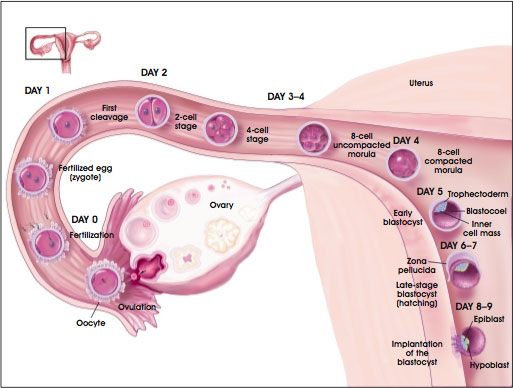
  • Day 0: fertilization takes place, usually in the upper fallopian tube
  • Day 3: the morula descends down the fallopian tube
  • Day 4: the morula enters the uterus
  • Day 5: the morula becomes a blastocyst
  • Day 6: the blastocyst begins to implant into the wall of the uterus
  • Day 10: implantation completes

This image, while somewhat detailed, depicts the timeline as the egg travels from the ovary, through the fallopian tube, to the uterus.


All of this assumes a normal sequence, beginning with ovulation and ending in implantation in the uterus.  When something goes awry, with can think of it as an ectopic implantation--not in the upper uterus.  In order of most common to least common:

  1. The fallopian tube: accounts for 95% of all ectopic pregnancies, it cannot grow like the uterus and will burst 
  2. Lower uterus/cervix: can be carried to term with careful following, placenta can implant over cervix and risks tearing
  3. Abdominal cavity: the fertilized egg will implant to live off of the mesenteric blood supply, can be carried to term but only deliverable through c-section
  4. The opposite fallopian tube
Ectopic pregnancies carry significant risk to the mother, since only the uterus serves for the place in which a baby should grow.  This does not mean that some ectopic pregnancies cannot end with delivery, but not all will have such an outcome.  The answer to the question of how to handle an ectopic pregnancy: carried to term vs. aborted, carries significant ethical and legal implications.  To anyone going into the field I strongly suggest an understanding of this topic.  

ResearchBlogging.org

Dickens, B. (2003). Ectopic pregnancy and emergency care: ethical and legal issues International Journal of Gynecology & Obstetrics, 82 (1), 121-126 DOI: 10.1016/S0020-7292(03)00175-9


Monday, March 14, 2011

Heme/Onc: understanding anemia

When admitted to the hospital almost all patients will receive a standard battery of blood tests, which includes a Complete Blood Count (CBC).  Often a primary care office will order such work for their patients simply to have a baseline, and many specialists will do so as a means to monitor drug reactions and overall health.  A CBC includes the measurement of many different components of blood, including the number of red blood cells, white blood cells, platelets, hemoglobin, hematocrit, and mean corpuscular volume.  The shorthand of the CBC looks like this:

On the left is the number of white blood cells; on the right- platelets; the H & H is in the middle, referring to the hemoglobin and hematocrit, with the hemoglobin on top.  For anemia the Hemoglobin (Hgb) matters most.  While it doesn't appear in the shorthand form of the CBC, the Mean Corpuscular Volume (MCV) also matters since it allows for an easy classification of anemia.  

Anemia results from a decreased oxygen delivery by the blood.  Symptoms can include many different manifestations including fatigue, lethargy, cold intolerance, and even passing out!  Anyone can address the symptoms, but only in knowing the type and cause of the anemia can one hope for an overall cure.  This does not mean that the anemia can't self resolve.  Many people may become transiently anemic yet never know it, but if seeking medical attention the patient will want as much information as possible.  

Mean Corpuscular Volume refers to the average red blood cell volume.  An anemia will very often affect this number thus allowing for a very simple classification, based on whether the number occurs within normal limits.  The normal value for an MCV ranges between 80 - 100.  The system of classification is as follows:
  • Normocytic: MCV between 80 - 100
  • Microcytic: MCV less than 80
  • Macrocytic: MCV greater than 100
Knowing the type of anemia starts the search for a cause, but in no way gives a definitive answer on its own.  But at least you can sound smart by beginning with a classification of the anemia.  

Microcytic anemias, those with an MCV below 80, often have red blood cells which look pale compared to normal--a phenomenon known as hypochromasia.  Numerous causes exist for a microcytic anemia, but keep in mind that while a microcytic state may point in one direction it is still important to consider causes which typically present as normocytic or macrocytic.  That said, the most typical causes include: 
  • Iron deficiency
  • Thalassemia
  • Sideroblastic anemia
  • Anemia of chronic disease
To understand the entities which compromise microcytic anemias requires an understanding of hemoglobin--the oxygen carrying compound in red blood cells.  Hemoglobin has two components:
  • Heme: the iron containing molecule of hemoglobin
  • Globin chains: four subunit chains defined as either alpha, beta, gamma, or delta
These components must come together correctly in order to have a properly functioning hemoglobin.  While iron deficiency affects heme, it is thalassemia which affects the globin chains.  Thalassemia results from a genetic mutation affecting the formation of the globin chains.  Normal adult hemoglobin has two alpha and two beta chains, and normal fetal hemoglobin has two alpha and two gamma chains; the condition is named for the chain it affects.  Thus alpha thalassemia affects the formation of the alpha chain, and so on.  

One must also understand sideroblastic anemia, a condition which results when the bone marrow makes ringed sideroblasts rather than typical red blood cells.  While it sounds confusing just understand that ringed sideroblasts are basically red blood cells which have nuclei as well as iron deposits.  Any number of things can cause a sideroblastic anemia, but four particular causes to know:
  • Alcohol
  • Pyridoxine deficiency
  • Lead poisoning
  • Myelodysplasia
A prussian blue stain, which will pick up the iron, may appear as follows:

Normocytic anemias occur within the normal limits of the MCV value.  Again keep in mind that causes between the different anemias may overlap:
  • Aplastic anemia
  • Sideroblastic anemia
  • Anemia of chronic disease
  • Fluid overload
Aplastic anemia occurs when the bone marrow does not respond adequately to the need for new RBC production.  To measure this look at the reticulocyte count, which is normally less than 2.  During times in which the bone marrow must make new RBCs the reticulocyte count should rise above 2, if it does not consider that the bone marrow has not appropriately responded to the stimulus for more production.  

Macrocytic anemias occur when the MCV has surpassed 100.  When thinking of this type of anemia think of diet induced.  Although not always true it covers the three most important causes:
  • Vitamin B12 deficiency
  • Folate deficiency
  • Alcohol
Thus comes the biggest question: how does one evaluate an anemia after looking at the MCV?  

Simple: order more tests!  

Absolutely standard tests for anemia should include a measure of serum iron, a ferritin level, and the calculated Total Iron Binding Capacity (TIBC), as well as a Serum Protein Electro Phoresis (SPEP), Urine Protein Electro Phoresis (UPEP), and a level of B12 and folate.  

Other tests to order may include a Lactate DeHydrogenase (LDH), which is released during cell destruction and serves as a particularly useful marker for RBC destruction.  

I can go into further detail if anyone would want that.  Just message me.  In the meantime I find an interesting article, which I have cited, in the AMA's Archives of Internal Medicine.  Written in 1958, it describes the clinical application of SPEP, something taken for granted today.  You may find it a waste of time to read, but it is interesting to see how science has progressed!

ResearchBlogging.org 
Robert Wall (1958). The Use of Serum Protein Electrophoresis in Clinical Medicine Archives of Internal Medicine, 618-658

Monday, March 7, 2011

The importance of the cardiac physical exam?

If it hasn't seemed apparent enough in the previous posts, I once again want to stress the importance of the physical exam.  By taking the time to thoroughly examine the patient, which includes a thorough history, tragic events like this can be avoided.

Numerous things should happen during any physical exam, but the cardiovascular exam should include: visual examination, palpation, and auscultation; with most of the focus on the neck and chest.

So what to do...

Take a few blood pressures.  Check each arm and one leg, normally the leg will have the higher blood pressure.  If not, add coarctation of the aorta to the differential.

Feel for pulses.  While weak distal pulses does not have to mean pathology, left ventricular hypertrophy is one possible cause.  Keep in mind that LVH occurs due to conditions such as AS, IHSS, AR, MR, and event HTN.

Look at the eyes.  Using the ophthalmoscope check for neovascularization, which suggests the blockage of arteries having resulted in the formation of new ones.  Two important disease entities to consider if you see this: diabetes, and coronary artery disease.

Examine the neck.  This means giving more than a shear glance and looking at the internal jugular vein, which includes checking for differences during inspiration and expiration; and listening for bruits in the carotid arteries.

  • Bruit: the turbulent flow of blood past an obstruction, otherwise understood as the abnormal sound blood makes as it passes an obstruction.  Because of the pumping of blood the bruit will make two sounds--the upstroke and the decline.  The importance lies in the speed of the upstroke and the speed of the decline, which together can aid in a diagnosis.
    • Rapid upstroke, Rapid decline: aortic regurgitation 
    • Rapid upstroke, Delayed decline: IHSS
    • Delayed upstroke, Delayed decline: aortic stenosis
    • Slow upstroke, Normal decline: atherosclerosis, only occurs in the carotid with the plaque
  • Internal Jugular Vein: correlates with pressure in the right atrium.  The pulse should normally be seen around 5 centimeters above the Angle of Louis of the sternum.  To look for this the patient should lay on the table at an incline of 30 to 45 degrees.  For comparison: 10cm from the Angle is the mandible; at 12cm is the ear lobe.  Two important entities need consideration when checking the internal jugular: 
    • Hepatojugular Reflux: a sign of right atrial overload, will stay elevated on inspiration and expiration.
    • Kussmual's Signs: distention of the internal jugular with inspiration; suggestive of pericardial effusion and impending respiratory failure.
Next, move on to the chest.

Palpate the chest.  Particularly check three areas:
  • Apex: should be at the left 5th intercostal space at the midclavicular line, but can vary slightly depending on the heart's deviation.  However, barely feeling it during S4 suggests a small heart, where as a large heart will generally create a sensation across all palpating fingers during S3.
  • Sternum: roughly located above the right ventricle.  Normally one will not feel it; only right ventricular hypertrophy is felt.  If felt you should add tricuspid regurgitation to your list, auscultating a systolic murmur in the tricuspid area would only further support this diagnosis.
  • Sternal border: correlates well with the left atrium, and just like the right ventricle it will only be felt if enlarged.  With left atrial enlargement consider disease states such as mitral stenosis, mitral regurgitation, and dilated cardiomyopathy.
Listen to the heart.  Listen for heart sounds and check for murmurs.  Focus on the four main areas:
  • Aortic: the right 2nd intercostal space
  • Pulmonic: the left 2nd intercostal space
  • Tricuspid: the left sternal border
  • Mitral: the left 5th intercostal space at the midclavicular line
And of course: always take a good history

More on heart sounds and cardiac history taking later...


Unfortunately the physical exam has lost its importance to many practitioners.  

In lieu of looking, listening, and palpating CT, MRI, US, and XR have come in as replacements.  While radiographic and ultrasonographic imaging have benefits, they have downsides as well.  After all, a physical exam doesn't expose the patient to radiation, require a contrast medium, or cost any more money than the salary to employ the practitioner.  But in a society clamoring for big money lawsuits clinicians need to protect themselves, and imaging modalities can provide conclusive answers without the subjectivity of the clinician's skills.  

Some medical schools require their students to do rotations in rural areas, which often lack the bells and whistles of large cities.  It is unfortunate that the bodies which govern medical education across the board don't mandate such training.  Only when one must go without reliance on imaging will he or she learn to appreciate a good physical exam.  

ResearchBlogging.org
Jauhar, S. (2006). The Demise of the Physical Exam New England Journal of Medicine, 354 (6), 548-551 DOI: 10.1056/NEJMp068013

Thursday, March 3, 2011

Cardiology: Murmurs!

The stethoscope has practically become the icon of healthcare.  To wear a stethoscope suggests that a person has some sort of clinical knowledge.  After all, why else would a person have a stethoscope on their person if they did not actually know how to use it?  A stethoscope amplifies sound to make very subtle noises audible to the human ear.  Thus the sounds of the hearts valves closing, the movement of air into and out of the lungs, the bowels doing their thing, and so on get recognized clinically with the use of a stethoscope.  Without an understanding of what the sounds actually mean such information proves useless, allowing the stethoscope to serve as nothing more than a mere piece of jewelry.

When putting the stethoscope to the chest one can look at the chest as having four separate areas in which to listen for heart sounds:
  • Aortic: the right second intercostal space adjacent the sternum
  • Pulmonic: the left second intercostal space adjacent the sternum
  • Tricuspid: the left third and fourth intercostal spaces adjacent the sternum
  • Mitral: the left fifth intercostal space in the mid-clavicular line, often the point of maximal intensity
The location of a heart sound allows for the listener to determine the significance of a murmur based on the clinical picture.  Some of the sickest patients may have a murmur which needn't be addressed while some of the healthiest patients can have murmurs which signify the presence of a silent killer.   This means that the clinician wields a heavy tool in his or her stethoscope.  

Just as a jugular venous pressure wave shows the cardiac cycle of systole and diastole, the stethoscope auscultation, or listening to the heart provides an audible representation of heart function.  Normally two sounds, or beats get heard: S1 and S2.  When someone describes the heart as making a lub dub noise, they are referring to S1 and S2.
  • S1 is the sound of the bicuspid and tricuspid valves closing during systole, the aortic and pulmonic valves should be open
  • S2 is the sound of the aortic and pulmonic valves closing during diastole, the mitral and tricuspid valves should be open
To learn mumurs, which usually sound like a whoosh instead of or after a beat, it's easiest to think of them in terms of whether they occur in systole or diastole.  Knowing their location helps to limit the differential.

Systolic murmurs are those which occur during systole, and can be further classified upon when the murmur actually occurs.

  • Early to mid systolic murmurs
    • Pulmonic Stenosis (PS)
    • Tricuspid Regurgitation (TR)
    • Atrial Septal Defect (ASD)
    • Idiopathic Hypertrophic Subaortic Stenosis (IHSS)
    • Mitral Regurgitation (MR)
  • Mid to late systolic murmurs
    • Mitral Valve Prolapse (MVP)
    • Aortic Stenosis (AS)
  • Holocystolic--occurring throughout all of systole
    • Tricuspid Regurgitation (TR)
    • Mitral Regurgitation (MR)
    • Ventricular Septal Defect (VSD)
Notice that TR and MR can occur as either early/mid systolic as well as pan-systolic.  The difference being that something acute occurs early, where as a chronic condition happens throughout.  

Diastolic murmurs also occur, albeit considerably less to know than the list of 10 systolic murmurs.  Often one auscultates these best in the neck, and can quantify them based on the sound they make: a rumble vs. a blow.
  • Diastolic Rumbles
    • Tricuspid Stenosis (TS)
    • Mitral Stenosis (MS)
    • Austin Flint 
  • Diastolic Blows
    • Aortic Regurgitation (AR)
    • Pulmonic Regurgitation (MR)
Once the clinician has auscultated a murmur he or she must somehow differentiate the type.  An echocardiogram can effectively diagnose problems with valves and septa (the plural for septum, not the transit authority).  However, since not always practical or available, the practitioner should remember a few fundamentals to accurately diagnose a murmur by physical exam.  Since the heart handles systemic circulation, look at the heart as a dual circuit: the right side of the heart receives blood from the venous circulation, anything which increases venous return will increase a murmur on the right; the left side of the heart pumps blood into arterial circulation, anything which increases the arterial blood pressure will increase a left sided murmur.  
  • Inspiration & leg raising: increase the amount of blood from venous circulation returning to the heart, this increase in preload leads to an increase in the duration of right sided murmurs
  • Squatting: increases the amount of blood returning to the heart as well as the systemic arterial blood pressure, this increase in both preload and afterload results in increased duration of right and left sided murmurs
  • Hand grip: constriction of the radial and ulnar arteries leads to a backup of arterial blood which increases the arterial blood pressure, this increase in afterload causes an increase in left sided murmurs
  • Valsalva & standing: leads to a decrease in both preload and afterload, ultimately reducing the duration of both right sided and left sided murmurs
One very important exception exists to these rules: IHSS
  • Duration increases with valsalva
  • Duration decreases with squatting
In considering the pathophysiology of IHSS, it makes sense.  Because of the hypertrophy of the subaortic area of the left ventricle blood will have a difficult time getting from the left ventricle into the aorta.  However, if the pressure in the arterial circulation increases it will lead to an increase in left ventricular pressure in order to compensate.  Where as this will increase the effect of most left-sided pathologies and thus increase their associated murmurs, in IHSS the pressure increase will push against the hypertrohpied tissue and essentially push it out of the way such that blood will move by easier and result in a diminished murmur.  Following the same logic the opposite is also true.  

Some people could not help themselves and insisted on putting their namesake to a diastolic murmur.  Nonetheless three particularly important ones exist:
  • Carey Coombs Murmur: the diastolic murmur of mitral stenosis heard as a result of rheumatic heart disease
  • Graham Steell Murmur: the diastolic murmur of pulmonic valve regurgitation
  • Austin Flint Murmur: the diastolic murmur heard best in the mitral area associated with severe aortic regurgitation compressing against a normal mitral valve

Although not very recent, a 1990 study showed that in diagnosing pediatric murmurs the clinical skills of an experienced cardiologist have a sensitivity of 96% and specificity of 95%.  While it may seem great, just ask yourself this: how many doctors today rely on their physical exam skills as much as those who practiced decades ago?  Just how many doctors today actually qualify as experienced as determined by that article?  Since the article left out such criteria I cannot know, but I suspect the number would be considerably less. 


ResearchBlogging.org

John F. Smythe MD, Otto H. P. Teixeira MD, Peter Vlad MD, Pierre Paul Demers MD, & William Feldman MD (1991). Initial evaluation of heart murmurs: Are laboratory tests necessary? The Indian Journal of Pediatrics, 58 (2), 231-231 DOI: 10.1007/BF02751126

Monday, February 28, 2011

Cardiology: Jugular Venous Pressure (JVP)

If you ask anyone with a clinical background to explain bradycardia, you will get a very simple yet sufficient response: a heart rate below 60 beats per minute (bpm).  However, if you ask that same person to relate it to jugular venous pressure, more often than not you can expect to receive a blank stare.  Why?  Simple: very few actually understand venous pressure waves. 

Jugular venous waveforms quantify the pressure within the venous system—which ultimately feeds into the right side of the heart.  Thus an abnormal waveform suggests the presence of pathology.  To understand an abnormality requires knowledge of the norm.  Without a concrete basis of what jugular venous pressure should be, one will struggle to identify a deviation from the standard.  

First, one must understand basic heart anatomy and function. 


The heart has four chambers.
  •  The Right Atrium, which receives deoxygenated blood from the superior vena cava and inferior vena cava and pumps its blood through the tricuspid valve (one of the atrioventricular valves) into the right ventricle
  •  The Right Ventricle, which receives blood from the right atrium and pumps it through the pulmonic valve and into the pulmonary artery towards the lungs
  • The Left Atrium, which receives blood from the lungs via the pulmonary vein and propels it through the mitral valve (AKA bicuspid valve, one of the atrioventricular valves) into the left ventricle
  • The Left Ventricle, which receives blood from the left atrium and pumps it through the aortic valve into the aorta for systemic circulation
The pumping of blood through the heart occurs in a two step process: diastole and systole, both of which can be thought of through their function in the ventricles.

  • Diastole happens first, in which blood fills the ventricles.  At first the low pressure in the empty ventricles drives the process but ultimately the atria contract to expel their contents into the ventricles
  • Systole happens next, in which the ventricles contract and push their contents into either the pulmonary artery or the aorta

With this very basic foundation of heart structure and function one can now look at the jugular venous pressure waveform, and keeping in mind that the internal jugular vein ultimately feeds into the superior vena cava one can understand how this waveform has clinical significance.


Each letter of the jugular venous pressure wave corresponds with a part of the heart’s normal cycle.
  •  A wave relates to atrial contraction, marking the end of diastole
  • C wave denotes the beginning of systole, at which point the pressure in the ventricles pushes against the atrioventricular valves which causes them to bulge ever so slightly
  •  X depression happens when the lower pressure in the atria allows for blood to fill in
  •  V wave occurs when the pressure in the atrium rises due to an increasing amount of blood along with a closed atrioventricular valve
  •  Y depression occurs when the atrioventricular valve opens and blood begins filling the ventricle right before atrial contraction

Just think of it in terms of blood in the vena cava: an A wave occurs because the pressure of the contracting atria puts resistance against the flow of blood out of the vena cava; and so on.  Thus with an understanding of the norm one can recognize pathology: any deviation from the curve above has significance.  It’s easiest to start at A and move from there:
  •  Large A wave: resulting from an increased pressure against which the atria contract
    • Seen with pulmonic and aortic stenosis and IHSS, seen with normal sinus rhythm in stenosis of the mitral and tricuspid valves, seen in ischemic heart disease, and can also occur due to aging
  • Canon A wave: large intermittent A waves which occur due to a problem with conduction in the heart which results in contraction of the right atrium against a closed tricuspid valve
    • Seen in arrhythmias, including paroxysmal atrial tachycardia and atrial flutter
  • Absent A wave: loss of A wave due to inability of atria to effectively contract
    • Seen with atrial fibrillation
  • Large V wave: due to an increased pressure in the atrium above normal
    • Seen with tricuspid regurgitation
  • Depression wave abnormalities
    • X descent: abnormality suggestive of acute pericardial effusion, such as in tamponade
    •  Y descent: abnormality suggestive of slow pericardial effusion, such as in chronic renal failure and with SLE

Of course all of this information hinges on one very important question: how does one assess the jugular venous pulse?  After all, what good does such data serve if one cannot apply it? 

To find the jugular venous pulse (jvp), the patient should be laying with their head elevated between 30-45 degrees̊.  After finding the pulsation of the internal jugular vein in the neck one should roughly measure from the jvp to the Angle of Louis—right above the sternum, which is typically around 5cm.  A significant deviation would suggest pathology. 

The reliability of the individual practitioner in finding the jugular venous pulse is notoriously unreliable.  Given the safety of ultrasound, its relatively cheap cost and its easy access, recent studies have looked into the possibility of ultrasound guided determination of the jugular venous pulse.  Two separate studies in 2010, one by Northern Ontario Medical School, and the other by the University of Iowa have suggested a much higher reliability in ultrasound-determined jugular venous pulse.  However, as with any new technological method it warrants further research before its application clinically.


ResearchBlogging.org

Socransky SJ, Wiss R, Robins R, Anawati A, Roy MA, & Yeung IC (2010). Defining normal jugular venous pressure with ultrasonography. CJEM : Canadian journal of emergency medical care = JCMU : journal canadien de soins medicaux d'urgence, 12 (4), 320-4 PMID: 20650024

Deol GR, Collett N, Ashby A, & Schmidt GA (2011). Ultrasound accurately reflects the jugular venous examination but underestimates central venous pressure. Chest, 139 (1), 95-100 PMID: 20798190

Sunday, October 17, 2010

Sugar Baker Procedure

In clinical science, if you ever hear of something with an unusual name, chances are it has something unique about it.  Consider an appendectomy--a boring name for the removal of the appendix, or cholecystectomy--a snooze of a term to describe the removal of the gallbladder; but both names serve a purpose in that they describe the procedure.  Now consider the Sugar Baker procedure.  Based on name alone, you don't have much to go by, unless you incorrectly suspect that it has something to do with confectionery. 

In talking with someone, they mentioned the Sugar Baker procedure, and it piqued my interest to the point that I wanted to know more about it.  What did it entail?  How did it get its name?  Why did one do it?  Thus, I went to the handy-dandy tool I often use--Google Scholar.  I found some helpful articles, but nothing of particular interest.  After resigning to the fact that I needed to do a more intensive search, I found a useful article: the treatment of peritoneal mesothelioma using the Sugar Baker procedure.  Along with a type of advanced stage colon cancer, these two cancers are the primary indications for the Sugar Baker procedure.

Despite scouring sources for information on peritoneal mesothelioma, I found little to describe this rare cancer's pathogenesis.  We have long associated asbestos as the primary cause of mesothelioma, but this applies when it develops in the lungs.  To date nobody has developed a concrete pathway for the development of mesothelioma in the peritoneum--the lining of the abdomen.  At least a third of patients with peritoneal mesothelioma have no history of asbestos exposure.  Without an understanding of how asbestos could get into the peritoneum exclusively, and a detailed social history of all patients to rule out asbestos exposure, the link between asbestos and peritoneal mesothelioma remains merely a working hypothesis.

Nonetheless, as with its pulmonary counterpart, it has a grim outcome.  Being rare doesn't help, since most money goes into the research of the more common malignancies.  As with all cancers, the only potential cure revolves around the possibility of the removal of all cancer cells.  To successfully rid the body of cancer, you must either completely remove or kill all cancer cells; typically done through surgery, radiology, or chemotherapy.  Each of these methods has its limitations; not all cancers respond to chemotherapy, some parts of the body can't take radiation, and sometimes surgery just isn't feasible.  In the case of peritoneal mesothelioma, surgery is the treatment of choice--the Sugar Baker procedure; only recently did surgeons come to recognize its efficacy.

Named after the physician who first employed it, the Sugar Baker procedure takes a lot of time and will have a heavy impact on the patient's life.  Basically, you cut open the abdomen, and remove all visible cancer.  Some things cannot be removed, but things that can come out include the colon, the spleen, the gallbladder, the omentum, and the peritoneum...as much as possible in so far as it is possible to remove the cancer.  After surgical removal of as much cancer as possible with regard to what must stay, the abdomen is then filled with heated chemotherapeutics, such as cisplatin or doxyrubicin; again, in the hopes of killing any remaining cancer. 

A 2010 study investigated the use of the Sugar Baker procedure for periteonal mesothelioma.  In looking at 20 patients, it found that in the years following the procedure, only six of the patients survived without disease recurrence.  Just over 25%, with further follow-up necessary to track the possibility of future cancer development. 

I have heard people debate the use of the Sugar Baker procedure, given its possible outcomes.  After all, living without a spleen and a colon does not come without consequence.  But then again, it gives the potential of life to someone otherwise destined for death.


ResearchBlogging.org Tudor EC, Chua TC, Liauw W, & Morris DL (2010). Risk factors and clinicopathological study of prognostic factors in the peritoneal mesothelioma. The American surgeon, 76 (4), 400-5 PMID: 20420251